
What is a Mammogram?
A mammogram is a specialised radiograph of the breast. It is a safe procedure with very low radiation used to show the breast tissue. Each breast is imaged separately. The breast is held gently but firmly between two plastic plates on the mammography machine for a few seconds.
Two x-ray views are taken when you undergo a breast screening mammogram: one downwards from the top of the breast and the other vertically from the side of the breast.


Types of Mammograms
Mammography is radiography or x-ray of the breast, and is referred to as either:
- Breast screening mammography - a wellness check using breast x-rays
- Breast diagnostic mammography - breast x-rays for a person with a breast concern: a lump, thickness and/or pain.
Screening mammography is a wellness test, and may detect breast problems before you have any symptoms or concerns. It may show changes in the breast up to two years before you or your doctor can feel a breast lump. This early detection of small breast cancers is important as it enables early treatment for the cancer.
Diagnostic mammography is used to assist in the diagnosis of an area of concern already identified by you or your doctor.
We are proud to be the 1st
Breast Care Clinic in Auckland to
offer 3D Breast Tomosynthesis.
What is a Three Dimensional (3D) mammogram?
A Three-dimensional (3D) mammogram, also known as breast tomosynthesis, is a type of digital mammography in which x-ray machines are used to take many pictures of thin slices of the breast to produce a 3D image. This process is similar to how a computed tomography (CT) scanner produces images of structures inside of the body.
- With a mammogram, glandular and fibrous tissue is white and fat is seen as black.
- Cancers are also seen as white so they can hide in normal glandular breast tissue, particularly if the breasts are dense which is seen in our clinic population.
- The 3D tomosynthesis takes images through the breast while the mammogram is taken, and the computer reconstructs this into 1mm slices through the breast.
- Our specialist radiologist reads this in conjunction with the state of the art 2D digital images.
- This improves cancer detection and reduces the number of unnecessary biopsies.
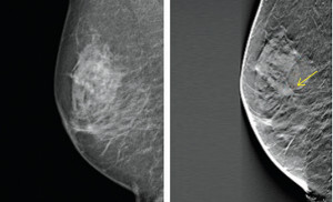
Cancer visible on the right 3D mammogram but not the left 2D mammogram.
Why are we using this technology?
- Increases the cancer detection rate
- Reduces false negative mammogram results especially in women with dense breast tissue
- Reduces the recall rate
- Reduces unnecessary biopsies
- Provides a 3D image to the surgeon
When should women undergo
mammography screening?
Regular screening mammography is recommended for all women over 40 years of age. Plus there are other recommendations:
-
Women from 40-50 years should have an annual mammography.
-
Women over 50 years of age may continue to have annual mammography or opt to have two-yearly mammography. If you are unsure which applies to you, discuss this with your doctor.
-
Women who have had a previous breast cancer should have an annual mammography.
-
Women who have a first degree relative (ie, their mother or a sister) who has had breast cancer should start having mammograms ten years in age before the age at which their relative was diagnosed.
We maintain strict processes to ensure our mammography service meets international quality standards.
Pre Examination
You will be asked to remove your clothing from the waist up, and given a gown to wear. For ease of undressing, you may like to wear a shirt and a skirt, or trousers.
Please do not wear deodorant or talcum powder on your breasts or armpits on the day of your examination. We do however, provide wipes and deodorant for your personal use.
Discuss any breast symptoms or problems with the mammographer before the examination and bring along any previous mammography films to your appointment.
It is important for the radiologists interpreting the mammogram to have previous studies for comparison as they compare previous films with new images to look for any areas of change to the breast.
Please remember to also bring the referral note from your doctor.
The Examination
A skilled mammographer will take at least two pictures of each breast. Each breast is placed on the x-ray machine and compressed with a plastic paddle. This flattening of the breast will reduce the breast thickness and hold the breast still, which optimises image quality and reduces radiation dosage.
Most women would describe the compression as uncomfortable, rather than painful. If you have found the compression painful, please discuss your concerns with the mammographer.
Sometimes further x-ray views may be needed to show an area of the breast more clearly. Don't be concerned if this occurs. An ultrasound scan may also be necessary to complete your examination.
In most instances having a mammogram only takes about 20 minutes.
We may in some cases also need to perform an ultrasound.
Post Examination
After the images are taken of the breast, you will be asked to wait until the mammographer has viewed them to see if any more are needed.
The Radiologist may ask if you wish to see the images and will give you your result then.